


Onychomycosis is a type of fungal lesion that exclusively affects the surface of the nails.To prevent the infection from spreading on time, you need to know what the nail fungus looks like, as the therapeutic effect will be achieved faster if treatment begins at an early stage of infection.Most often, the disease is found in the elderly due to poor immunity, but the risk of getting a disease refers to every person.There is no single classification of nail fungal damage;In medical practice, it is customary to distinguish them in place of localization and the depth to which they penetrate.The infection is also grouped depending on the type of pathogen.
Types of pathogens and primary characters
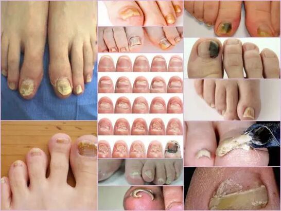
The symptoms of nail fungus in the initial stage are easily determined directly by the condition of the nail.This sign is most information, since onychomycosis always manifests itself in the form of changes in surface color, deformation of the bed, exfoliation and any external changes.The latter are expressed by roughness, the formation of channels and cracks, as well as point crossed and general nail disorder.
The main sign of a healthy nail is pink and transparency.Onychomycosis at each stage is characterized by the turbidity of the nail and the color change of yellow, brownish or greenish-black.At an advanced stage, the surface can acquire a black shade against the background of almost complete destruction.
The external signs of a fungus infection depend on the type of pathogen that becomes infected.The following possible lesions are distinguished in medical practice:
- Candida infection with a fungus, which is expressed by the release of the nail directly at the base of the box.Candidiasis of the rollers around the nail will be characteristic of candidiasis.This version of the complication may either have a bacterial source in the form of streptococci or staphylococci, or be expressed in the form of medium or psoriasis plaques;
- Rubrum trichophyton type dermatophytes.In this case, the penetration of the infection occurs directly from the free edge of the nail.The first symptoms of such a pathogen are the appearance of a yellowish spot on the surface.In the neoplasm field, the nail breaks down, and the spot itself tends to increase.The common place for localization of the neoplasm is along the plaque, parallel to the nail rollers, in which case the infection is called distal-based.There is another form of damage from this pathogen - distal, in which the stain appears from the side removed from the hole, mainly in the middle of the free edge;
Important!
This type of fungus is most commonly found on the toe, gradually turning the nails into a free yellowish mass.In the absence of proper treatment, the disease goes into hyperkeratosis.The nails are completely destroyed due to the spread of infection around the perimeter.
- Dermatophytes such as trichophytone mentalities.Onychomycosis with such arousal is also most often based on the nails of the thumbs, less than the small fingers.An infection with a fungus of this type requires therapy not only of the nail but also the legs due to the rapid spread of the pathogen.Symptomatic disease can be like leikonichia, a disease common in medical practice.The main signs are white spots that appear in different parts of the nail, neoplasms are distinguished by non -standard shape and different sizes.It is easy to distinguish the fungus from Leakonichia - in the latter case spots are the accumulation of air, which is not observed with damage to the fungi;
- Fungus of mildew.This damage option is significantly less than the applicant or dermatophytic form.The main sign of just such an infection - the surface of the plate acquires a dark, almost black shade.Not the whole finger can be completely infected, but only part of the nails.The first signs of leg fungus of this type is a sudden change in color.Onychomycosis can develop in the form of a longitudinal strip of black or dark green in the background of the rest of the pink part of the nail.
Diagnostics by type of change
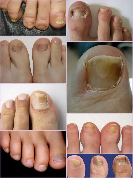
It is not difficult to notice a fungus of the toenails even at the primary stages of infection, as infection of this type is very active from the first day of the lesion.Instead of the usual transparent nails of pale pink in the patient, significant surface deformation and general change in condition are observed.The affected area has a matte dull yellowish shade that appears mainly on the thumbs.The type of fungi and the degree of damage are the determining factors.
In the first stage, the fungal damage to the legs on the legs is the appearance of small foci.The thickening of the plate and keratinization of the bed under the nail will be characteristic.This stage is accompanied by such a phenomenon as partial detachment and ejection of the nails, which serves as a source of infection for healthy people.
Despite the active thickening of the plate, its constant digestion, regardless of the current factors, can be observed.The characteristics of each stage and the symptoms of the fungus on the legs depend directly on the type of pathogen.
Depending on the changes occurring with the nails, there are three options for onychomycosis:
- Hypertrophic;
- Normotrophic;
- Atrophic.
In the first case, there is a sudden change in the shade of the nails, its destruction at the edges, as well as the deformation of the surface of the plate.The nails thicken so much that it causes discomfort and painful sensations when walking.Normotrophic mycosis of the toenails is characterized by the presence of a healthy shine, but the dish itself acquires some jaundice, with spots forms on it.With an atrophic type of damage, brown and gray foci are formed on the surface of the nails.Thanks to them, it is possible to accurately determine the location of the pathogen.
Important!
The atrophic or onycholithic type of nail fungus is characterized by the thinning of the nail plate, not its growth, as in other cases.Those areas where the pathogen is located are inclined to separate.The lack of appropriate treatment leads to an advanced stage - complete rejection of the nails.
Location
Another sign through which you can release fungal damage to your toenails depends directly on the location of the foci on the nails.This also includes the depth of the pathogen, which in turn allows you to determine the approximate duration of upcoming therapy and the chances of rapid recovery.
The fungal diseases of the toenails at the location site are classified into the following groups:
- Onychomycosis is a type of white surface - the appearance of many whitish spots on the surface of the nails.The fungal infection leads to the tear of the skin at the sites of the appearance of spots, which are actively disposing of flakes.The advanced stage leads to complete destruction and rejection of the plate;
- Distal - develops on the free edge of the nail.The color change is observed first in a small area of the plate, after which there is an active expansion of its borders.The lesion is characterized by a yellowish-gray or brown shade, as well as a rough uneven surface and gradual exfoliation;
- Side onychomycosis has the stages of development similar to distal, but it is located exclusively on the sides of the nails;
- General infection - complete infection on the entire surface;
- Proximal onychomycosis.This disease begins its activity from the cuticle, which becomes inflamed, then the fungus quickly affects the nail, and the process itself begins with the appearance of a small white spot near the inflammatory area of the rake roller.
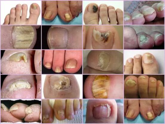
The most common forms of mycosis of the toenails are lateral and distal, which is usually caused by dermatophytes.Such forms of damage as proximal and white can act as a secondary disease that accompanies the disease of the immune system, for example, Vich.The overall damage to the nail with a fungus should be considered as an advanced stage of development of each fungus under the nail.
Fungus characteristics under the microscope
Despite the presence of an impressive number of signs of onychomycosis, other diseases associated with skin problems rather than infected by nature are considered to be fungal damage to the toenails.You can determine the exact diagnosis and the type of pathogen only in the laboratory under a microscope, for which the hospital is scraped by biological material from the affected areas.
The resulting biomaterial is pre -placed in an alkaline environment, after which a repeated increase is performed.The study of the nail fungus under a microscope allows you to see an active pathogen, the outer form of which determines its type, scale of distribution and approximate degree of damage.In the nutrient medium, you can predict the approximate growth of the colonies, which not only allows you to make an accurate diagnosis, but also to determine the limitation of the infection.
Important!
As it is possible to determine the presence of onychomycosis only in a laboratory, you should not delay a visit to the doctor with the less suspicion.The nail fungus develops rapidly and the loss of time increases therapy.
What is an alarming signal?
The fungus of the toenails is manifested by certain symptoms that are partially similar to some skin diseases.The following signs are more likely to show an onychomycotic lesion that requires medical intervention:
- The appearance of yellow spots on the plate, its deformation, a change in the structure that is not observed earlier;
- Darkening the plate, loss of transparency, some pictures of the nail sponge show a nuance close to black, which is characteristic of pathogens;
- Thickening and keratinization of the nails or back too acute thinning;
- Rejection of the nail from the bed, the tear of flakes and crumbs;
- Swelling of the roller, suspension over the plate, release of fluid;
- The nails affected by the fungus look inflamed, regardless of the type of pathogen.
All these symptoms show a high risk of infection.Some external symptoms of changing the nail structure can be the result of other diseases.With increased British, the amount of calcium and iron in the body should increase, increased tuberosis means any infection of the body, with pure white stripes and dots, lack of honey or zinc is possible.Despite the fact that in the photo, the nail fungus looks like a focal damage to the plate with a color change, closer to yellow, this does not always show the infection.Desire can show various problems with the stomach and liver.
It is not difficult to recognize onychomycosis of the fingers through external signs, despite the extensive classification of the disease.But you can determine the exact type of pathogen and the stage of damage can only be in the laboratory.